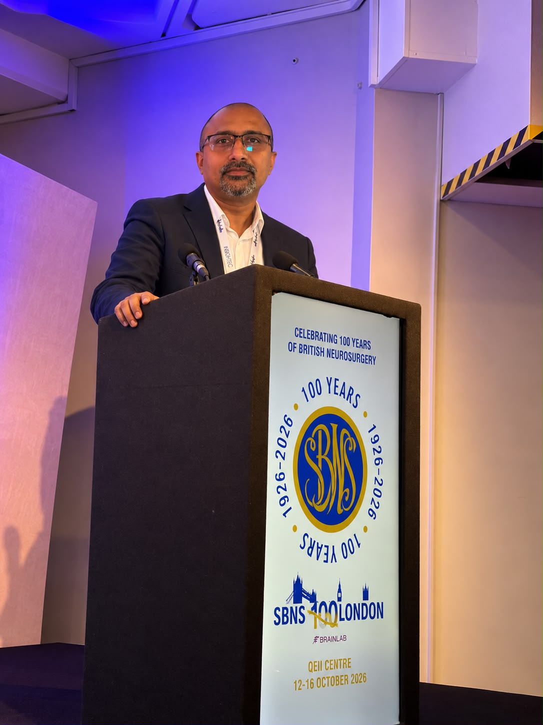

A 14-year-old regained the ability to walk after salvage surgery by Dr. Mohana Rao Patibandla, Guntur neurosurgeon, with five-year follow-up.
GUNTUR, ANDHRA PRADESH, INDIA, October 28, 2026 /EINPresswire.com/ — From Wheelchair-Bound to Walking Again: Five-Year Follow-Up Highlights Complex Spine Surgery Research by Guntur Neurosurgeon in London
A six-patient study presented at the Society of British Neurological Surgeons’ centenary meeting examines an important challenge in craniovertebral junction surgery: persistent pressure on the brainstem and spinal cord despite previous stabilisation.
A 14-year-old patient who gradually deteriorated to being wheelchair-bound despite an earlier spinal stabilisation procedure regained the ability to walk after further surgery and continues to do well five years later. The case highlights an important clinical challenge in complex neurosurgery: stabilising the spine does not always mean that pressure on the brainstem and spinal cord has been adequately relieved.
The case forms part of a six-patient clinical series presented by Dr. Mohana Rao Patibandla, Founder and Chief Neurosurgeon of Dr. Rao’s Hospital – International Institute of Neurosciences (IIN), Guntur, at the Society of British Neurological Surgeons (SBNS) centenary meeting in London in October 2026.
The presentation, titled “Transnasal Endoscopic Odontoidectomy for Failed Posterior Craniovertebral Fusion: Salvage Decompression and Outcomes in 6 Complex Cases,” examined the use of endoscopic surgery through the nasal passages to relieve persistent pressure at the junction of the skull and upper spine.
The central message is relevant to patients and families facing complex brainstem and upper-spine disorders: when neurological function continues to deteriorate after stabilisation surgery, specialists may need to reassess whether pressure on the nervous system remains.
A young patient’s journey from initial improvement to renewed walking
At 14 years of age, the patient underwent a DCER procedure at AIIMS, New Delhi, for a complex craniovertebral junction disorder. The patient initially improved following surgery. Over time, however, neurological function gradually deteriorated, eventually leaving the patient wheelchair-bound.
The clinical problem required a fresh assessment of the structures around the brainstem and upper spinal cord. In selected patients, abnormal bone or associated tissue can continue to exert pressure from the front, even after an operation intended to stabilise the skull–spine junction.
The patient subsequently underwent endoscopic transnasal odontoidectomy, a procedure in which surgeons use an endoscope and specialised instruments passed through the nose to reach the odontoid process, a bony projection of the second cervical vertebra. The aim is to remove the compressive bone and create more space for the brainstem and upper spinal cord.
Following the procedure, the patient regained the ability to walk. At five-year follow-up, the patient continues to do well, with the ability to walk maintained.
The case offers an encouraging example of sustained functional improvement after carefully selected salvage decompression. It also demonstrates why a patient’s neurological progress must be assessed alongside the stability of the spine.
The outcome is specific to this patient and does not guarantee a similar result for others. Treatment decisions depend on the underlying disorder, the location and severity of compression, previous operations and the patient’s neurological condition.
Why stabilising the spine may not be enough
The craniovertebral junction is the area where the skull connects to the upper cervical spine. It protects the transition between the brainstem and spinal cord, structures responsible for essential neurological functions.
In conditions such as basilar invagination, part of the upper spine projects abnormally towards the base of the skull. Another cause of compression is retro-odontoid pannus, abnormal tissue that forms behind the odontoid process and can press against the nearby neural structures.
Surgery from the back of the neck may help restore alignment and stabilise the bones. However, stabilisation and decompression are different objectives. A stable surgical construct does not, by itself, prove that pressure from the front has been relieved.
When neurological deterioration continues, doctors may need to review the patient’s symptoms, neurological examination and imaging to determine whether residual compression, instability or another problem is responsible.
The question is not simply whether the previous operation has stabilised the spine. It is whether the brainstem and spinal cord have adequate space and whether the underlying cause of neurological impairment has been addressed.
What did the six-patient study examine?
The clinical series included six patients treated between 2019 and 2022. Two patients had basilar invagination, while four had retro-odontoid pannus. The patients had different underlying conditions and surgical histories, making individual assessment essential.
The research examined endoscopic transnasal odontoidectomy as a salvage strategy for selected patients with persistent pressure at the front of the craniovertebral junction.
Neurological function was assessed using the Nurick grading scale, which clinicians use to describe the severity of disability associated with spinal cord dysfunction, particularly difficulties with walking.
In the study’s working dataset, the mean preoperative Nurick grade was 4.33, compared with a mean postoperative grade of 2.83. This represents a mean improvement of 1.50 Nurick grades.
These figures describe the average change across the group. They do not mean that every patient recovered completely or experienced the same degree of improvement. The five-year clinical course of the 14-year-old patient is a particularly notable individual outcome within the broader clinical experience.
Because the study involved only six patients and had no comparison group, its findings cannot establish that the procedure is superior to other surgical approaches. Larger studies and longer-term assessment across more patients are needed to better understand which individuals are most likely to benefit.
How does endoscopic transnasal odontoidectomy work?
The endoscopic transnasal approach allows surgeons to reach selected abnormalities at the front of the craniovertebral junction through the natural passages of the nose.
Using an endoscope for visualisation, the surgical team works towards the odontoid process and removes the portion of bone contributing to compression when indicated. The objective is to relieve pressure on the brainstem and upper spinal cord while carefully protecting nearby blood vessels, nerves and other delicate structures.
The procedure requires detailed planning based on CT and MRI findings, the patient’s anatomy and the previous surgical history. The team must also determine how stability will be maintained after decompression.
It is not a routine procedure for everyone who has undergone posterior fixation. Its potential role is limited to carefully selected patients in whom clinically significant ventral compression remains and the approach is considered appropriate.
The essential principle is to select the surgical route according to the pathology that needs to be treated, rather than assuming that the same approach is suitable for every patient.
What should patients and families take away from this research?
For patients with complex upper-spine disorders, three treatment goals need to be considered separately.
Stability: keeping the bones at the skull–spine junction in an appropriate and secure position.
Decompression: relieving pressure on the brainstem and spinal cord.
Neurological recovery: monitoring changes in walking, strength, balance and other affected functions.
These goals are closely related, but achieving one does not automatically guarantee the others.
If a patient continues to deteriorate or experiences persistent neurological problems after surgery, the appropriate response is specialist reassessment—not an automatic assumption that another operation is required. Clinical examination and imaging help determine the cause and guide further decisions.
The young patient’s recovery illustrates why reassessment can matter in selected complex cases. It also underlines the importance of long-term follow-up to understand whether improvement is maintained.
Research presented at an international neurosurgical meeting
Dr. Patibandla presented the six-patient series at the Society of British Neurological Surgeons’ centenary meeting in London in October 2026. The presentation brought experience from a specialised neurosurgical centre in Guntur into an international discussion of complex craniovertebral junction disorders and revision surgery.
The research highlights a difficult but important clinical problem: persistent compression may require a different surgical strategy from the one initially used to stabilise the spine. It also emphasises that decisions must be based on the individual patient’s anatomy, neurological condition and previous treatment.
The six cases provide clinical experience rather than definitive comparative evidence. Further collaborative research will be important in evaluating long-term outcomes, potential complications and the patients most likely to benefit from this approach.
About Dr. Mohana Rao Patibandla
Dr. Mohana Rao Patibandla is the Founder and Chief Neurosurgeon of Dr. Rao’s Hospital – International Institute of Neurosciences (IIN), Guntur, Andhra Pradesh, India. His areas of practice include complex brain and spine surgery, minimally invasive neurosurgery, endoscopic skull base surgery, cerebrovascular and endovascular neurosurgery, paediatric neurosurgery, neuro-oncology and stereotactic radiosurgery.
About Dr. Rao’s Hospital – International Institute of Neurosciences
Dr. Rao’s Hospital – International Institute of Neurosciences (IIN), based in Guntur, Andhra Pradesh, India, provides specialised evaluation and treatment for disorders of the brain, spine, nerves and cerebrovascular system. Its services include neurosurgery, spine surgery, endoscopic procedures and neurocritical care.
Media Contact
Dr. Rao’s Hospital – International Institute of Neurosciences (IIN)
Guntur, Andhra Pradesh, India
Phone: +91 90100 56444
Email: info@drraoshospitals.com
Website: https://drraoshospitals.com
Keywords: craniovertebral junction surgery, endoscopic transnasal odontoidectomy, basilar invagination, brainstem compression, spinal cord compression, posterior craniovertebral fusion, minimally invasive neurosurgery, spine surgery, Dr. Mohana Rao Patibandla, Guntur neurosurgeon
Mohana Rao Patibandla
Patibandla Narayana Swamy Neurosciences LLP
+91 90100 56444
info@drraoshospitals.com
Visit us on social media:
LinkedIn
Instagram
Facebook
YouTube
X
Dr. Rao’s Hosptial – the best neurosurgery hospital in India – with medical tourism
Legal Disclaimer:
EIN Presswire provides this news content “as is” without warranty of any kind. We do not accept any responsibility or liability
for the accuracy, content, images, videos, licenses, completeness, legality, or reliability of the information contained in this
article. If you have any complaints or copyright issues related to this article, kindly contact the author above.

Media gallery