
Reliable primary cell resources advance mechanistic exploration, drug screening and pre-clinical modelling for skin-mucosa research
HUSTON, TX, UNITED STATES, November 5, 2026 /EINPresswire.com/ — High-purity, high-viability, functionally stable primary cells with consistent batch-to-batch performance serve as the critical starting point for reliable experimental data and efficient project progression in skin physiological mechanism dissection, wound-repair pathway discovery, pathogenic gene validation, and screening evaluation of new drugs & functional cosmetic ingredients. Directly isolated from living tissues, primary cells retain native physiological features, differentiation potential and signal-response profiles, making them irreplaceable research tools for skin and mucosal-related studies. Nevertheless, primary cells impose stringent requirements on tissue harvesting, isolation workflows and culturing conditions. Any procedural defect may trigger compromised cell purity, reduced viability or microbial contamination, further undermining experimental reproducibility.
As an established player in life-science reagent development, Cloud-Clone leverages in-house SPF-grade animal housing facilities and a full-chain quality-control framework to launch more than 40 types of primary cells covering skin and mucosal tissues. These cell products deliver robust research support for studies on skin trauma, inflammation, barrier dysfunction, scar fibrosis and skin neoplasms.
1. Rigorous Qualification: Full-Chain Quality Control From Origin to Product Delivery
High-impact scientific outputs build upon strict control over raw biological materials. The quality of primary cells heavily depends on controllable tissue sources. Cloud-Clone operates three in-house SPF-grade animal facilities with a total area of approximately 1,000 m², equipped with standardized IVC housing systems. The company independently manages the complete workflow spanning animal breeding, husbandry and tissue collection, effectively mitigating batch variation introduced by external third-party suppliers.
For compliance credentials, Cloud-Clone holds ISO certifications for quality management, environmental management and occupational health & safety management. It also possesses official permits for experimental animal production and experimental animal utilization. Stringent source-level quality control and standardized husbandry guarantee high purity, high viability and contamination-free status for primary cell products, laying a solid and dependable foundation for your research assays.
2. Comprehensive Product Portfolio: Skin-Focused Primary Cell Library Across Multiple Species & Tissues
Cloud-Clone’s catalogue includes over 560 types of primary cells derived from more than 10 species including human, mouse, rat, rabbit, dog, cat, pig, sheep and guinea pig. Tailored for skin and mucosal research workflows, over 40 specialized primary cell products are available, covering epidermal, dermal, skin-appendage, cutaneous and luminal mucosal tissues:
– Epidermal cells: Adult / neonatal epidermal keratinocytes (AEK/NEK), oral keratinocytes (OKs), lingual epidermal cells (LiEC), and others.
– Dermal cells: Adult / foetal / neonatal dermal fibroblasts (ADF/FDF/NDF), dermal microvascular endothelial cells (DMEC).
– Adipose & skin-appendage-related cells: Subcutaneous preadipocytes (SPAD), visceral preadipocytes (VPA), adipose microvascular endothelial cells (AMEC).
These cell products are available in ready-to-ship stock. They are well-suited for skin physiological mechanism dissection, pathological model construction and new-drug screening assessment.
3. Top-Journal Case Validation: Robust Performance of Cloud-Clone Primary Cells in Complex Pathological Models
High-grade primary cell products must not only withstand routine laboratory assays but also satisfy stringent standards for high-profile research work.
In September 2025, a study published in Nature Communications (2025 IF = 18.1, JCR Q1) drew wide attention within regenerative medicine and metabolic disorder research communities. Research teams from the Chinese Academy of Sciences South China Sea Institute of Oceanology, Sun Yat-sen University and other institutes isolated a marine actinobacterium from deep-sea sediment collected at 3412 m depth in the Indian Ocean and identified polydecalinmycin (PDM), a structurally novel polyether ionophore featuring an octahydronaphthalene scaffold.
This work uncovered a dual-phase regulatory mechanism driving wound healing. In the acute phase, PDM triggers mitochondrial Na⁺ influx to induce transient ROS bursts and kick-start repair signalling cascades. Upon sustained exposure, PDM activates Nrf2-driven antioxidant responses to shield cells against high-glucose-provoked oxidative stress. Such “pro-repair first, anti-damage subsequently” functional profile enables PDM to exert superior therapeutic efficacy compared with EGF in diabetic non-healing wound models.
Within this complete evidence chain linking deep-sea microbial discovery to skin-repair evaluation, Cloud-Clone primary mouse dermal fibroblasts (PMDF, Cat.No. CSI088Mu01) served as one of the key skin-repair cellular models for verifying the pleiotropic pro-healing functions of PDM. This top-publication case validates the high reliability of Cloud-Clone primary cells in complex pathological modelling and demonstrates the value of our cell resources in supporting landmark scientific advances.
4. Visual Proof: Microscopic Validation for High Purity and Viability
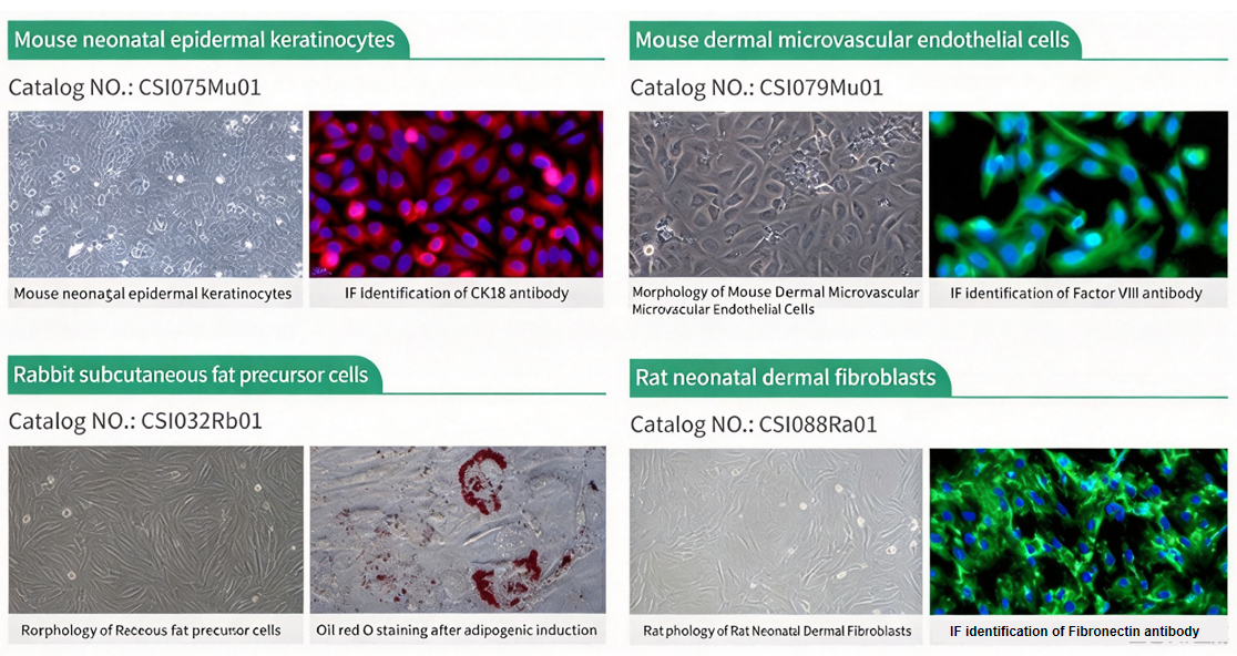
High-quality primary cells require well-document source information as well as verifiable identity confirmation. Every batch of Cloud-Clone primary cells undergoes morphological observation and immunofluorescence (IF) identification to guarantee homogeneous cell purity and differentiation status:
– Rabbit subcutaneous preadipocytes (CSI032Rb01): Distinct lipid droplets observable following adipogenic induction validated via Oil Red O staining.
– Canine adult epidermal keratinocytes (CSI073Ca01): Typical cobblestone morphology; positive signal from CK18 antibody immunofluorescence assay.
– Mouse dermal microvascular endothelial cells (CSI079Mu01): Representative spindle-to-polygonal morphology; positive identification using Factor VIII antibody.
– Feline adult dermal fibroblasts (CSI087Fe01) & Rat neonatal dermal fibroblasts (CSI088Ra01): Spindle-shaped morphology; positive signal from Fibronectin antibody identification.
– Rabbit lingual epidermal cells (CSI294Rb01): Excellent cell purity confirmed via CK18 antibody labelling.
Figure 1 Cloud-Clone primary cell products from skin and mucous membrane tissues
5. Application Portfolio: Empowering End-to-End Skin-Science Research
Cloud-Clone skin-series primary cells support diverse core research directions for skin and mucosal studies. The table below illustrates research objectives, applicable cell types and experimental values:
5.1 Skin Trauma and Wound-Healing Research
Skin wound healing relies on finely-orchestrated crosstalk across inflammatory, proliferative and remodelling phases. Keratinocytes, fibroblasts and endothelial cells execute distinct functional roles in this process. Epidermal keratinocytes (AEK/NEK) support investigations on re-epithelialisation-associated migration and proliferation mechanisms. Dermal fibroblasts (ADF/NDF) constitute core cellular readouts for collagen synthesis, granulation-tissue formation and contractility assessment (as exemplified in the aforementioned Nature Communications study). Dermal microvascular endothelial cells (DMEC) are suitable for angiogenesis and microcirculation reconstruction research. These cell types can be utilised individually or in co-culture setups to build complete research systems spanning epidermal re-coverage to dermal repair.
5.2 Skin Inflammation and Immune-Regulation Research
Dermal microvascular endothelial cells form the primary cellular barrier mediating leukocyte adhesion and trans-endothelial migration during cutaneous inflammatory episodes. DMEC and adipose microvascular endothelial cells (AMEC can be deployed to explore adhesion-molecule expression, barrier permeability shifts and endothelial-activation signalling upon inflammatory-cytokine stimulation. Combined with inflammatory-cytokine-secretion profiling of keratinocytes, researchers can construct in-vitro skin inflammatory microenvironments for anti-inflammatory active-ingredient screening and mechanistic validation.
5.3 Mucosal Injury and Repair Research
Oral and lingual mucosal tissues possess unique research value for wound-repair investigation and local-drug-irritation assessment. Oral keratinocytes (OKs) and lingual epidermal cells (LiEC) recapitulate mucosal-epithelial barrier physiology and regenerative responses. These cells fit well for mechanistic studies of mucosal injury repair, safety evaluation for topically-administered agents and mucosal-inflammation model construction. Multiple species including human, rabbit, dog, cat, sheep, mouse and rat are offered to satisfy species-matching requirements of diverse experimental frameworks.
5.4 Skin Barrier Deficiency and Scar Fibrosis Research
Adult and neonatal epidermal keratinocytes support evaluation of differentiation-marker expression, lipid biosynthesis and barrier-related gene regulation, applicable for barrier-damage mechanistic studies and in-vitro assessment of moisturising & skin-repair functional ingredients. Adult, foetal and neonatal dermal fibroblasts (ADF/FDF/NDF) serve as ideal cellular systems for studying fibrotic phenotypic switching, myofibroblast differentiation and anti-fibrotic intervention testing. Side-by-side comparison between foetal-derived and adult-derived fibroblasts facilitates exploration of mechanistic disparities between scar-free regeneration and fibrotic wound healing.
5.5 Skin Neoplasm and Adipose-Tissue Microenvironment Research
Subcutaneous preadipocytes (SPAD) and visceral preadipocytes (VPA) enable investigations into adipose-tissue functions within skin-tumour microenvironments, metabolic regulation and skin-appendage-associated disorders. After adipogenic induction, preadipocytes mature into adipocytes for lipid-metabolism research, obesity-linked cutaneous pathology and adipose-graft-relevant studies.
5.6 Pathological Model Construction & New-Drug / Functional-Ingredient Screening
Combined deployment of these primary-cell resources enables assembly of diverse in-vitro evaluation platforms for drug cytotoxicity profiling, high-throughput screening of functional raw materials, and alternative in-vitro assays for skin irritation and sensitisation assessment. High cell purity, stable viability and consistent differentiation status significantly improve screening reproducibility and translational potential.
As skin-mucosa-oriented research continuously evolves, access to biologically authentic primary cell resources remains indispensable for translating laboratory discoveries toward therapeutic applications. Cloud-Clone will keep expanding its primary-cell product catalogue, optimising full-chain quality-control protocols and collaborating with global research communities to accelerate breakthroughs in skin disease mechanism research, therapeutic-agent development and translational biomedicine. For detailed product specifications, batch-related documentation or customised technical support, please reach out to our global technical-support team.
About Cloud-Clone Corp.
Cloud-Clone Corp. is dedicated to the development and production of high-quality immunoassay reagents and detection solutions. With a focus on antibody engineering, multiplex assay development, and cross-platform compatibility, the company provides research tools designed to support precision medicine and advanced biomedical investigation globally. Our core products and services include the research and development of proteins, antibodies, ELISA kits, primary cells, and multiplex cytokine assay kits, as well as professional CRO services to fully meet the diverse needs of biomedical research and related fields.
For more information about Cloud-Clone Corp, visit www.cloud-clone.com.
CLOUD-CLONE CORP.(CCC)
Tel: 001-832-538-0970, 0086-27-8425-9552
Email: mail@cloud-clone.com, sales@cloud-clone.us
Visit us on social media:
LinkedIn: https://www.linkedin.com/company/cloud-clone-corp/
Instagram: https://www.instagram.com/cloudclonecorp2023/
TickTok: https://www.tiktok.com/@cloudclonecorp2026
SuKi Duan
CLOUD-CLONE CORP.WUHAN
Visit us on social media:
LinkedIn
Instagram
TikTok
X
Legal Disclaimer:
EIN Presswire provides this news content “as is” without warranty of any kind. We do not accept any responsibility or liability
for the accuracy, content, images, videos, licenses, completeness, legality, or reliability of the information contained in this
article. If you have any complaints or copyright issues related to this article, kindly contact the author above.

Media gallery